In a remarkable leap forward for paleontological research, a team of scientists from the Nanjing Institute of Geology and Palaeontology, Chinese Academy of Sciences, have developed a novel approach to analyze the three-dimensional structure and chemical composition of Ediacaran embryo-like fossils. These ancient microfossils, dating back to 590–570 million years ago, have long puzzled scientists due to their enigmatic affinities and the challenges associated with studying their delicate structures.
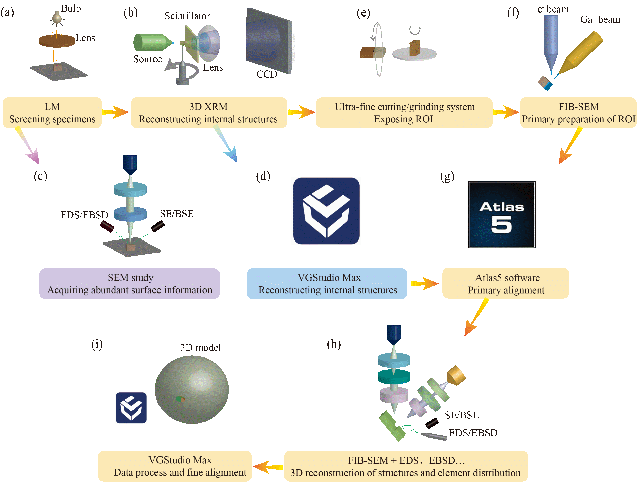
The study, published in the Journal of Earth Science, introduces an integrated method that harnesses the power of 3D X-ray microscopy (3D-XRM) and focused ion beam scanning electron microscopy (FIB-SEM). This cutting-edge technique allows researchers to peer into the microscopic intricacies of fossils with unprecedented clarity, offering a dual advantage: the non-destructive 3D visualization capabilities of 3D-XRM and the nanoscale chemical and structural analysis prowess of FIB-SEM.
Traditional fossil analysis techniques have reached their limits, struggling to provide the detailed, in-depth information required to understand the complex structures and chemical compositions of ancient organisms. The Ediacaran embryo-like fossils, in particular, with their spherical structures reminiscent of animal embryos, demanded a more sophisticated approach.
The researchers' innovative strategy involves a two-pronged attack: first, using 3D-XRM to create a non-destructive, high-resolution 3D model of the fossils, and then employing FIB-SEM to uncover the structural and chemical composition at the nanometer scale. This synergy between the two techniques overcomes the individual limitations of each, providing a comprehensive view of the fossils' structure and chemistry.
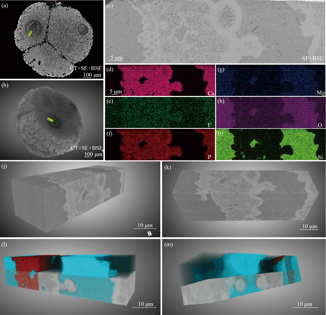
Through this advanced imaging technique, the team has successfully reconstructed the intricate, multi-layered structures within the cell nuclei of the Ediacaran embryo-like fossils. They discovered a concentric ring structure composed of two distinct mineral facies: one rich in clay minerals and the other in apatite, a form of calcium phosphate. This finding sheds new light on the preservation processes of these ancient organisms and their subcellular structures.
The ability to distinguish between biological structures and those formed through geological processes is a game-changer for paleobiologists. This research not only enhances our understanding of early life on Earth but also refines our ability to interpret the fossil record, offering insights into the evolutionary history of life.
The combined use of 3D-XRM and FIB-SEM is expected to open new avenues in paleontological and geological research. This method has the potential to transform the study of not only Ediacaran embryo-like fossils but also a wide range of geological specimens, from understanding the formation of minerals to exploring the origins of life on our planet.

Workflow diagram of the combined 3D-XRM and FIB-SEM technology method

The 3D structure and elemental composition of the cell nuclei in the embryo-like fossils from the Ediacaran Weng'an Biota
Download:
