
In a recent report published in National Science Review, a Chinese team of scientists highlights the discovery of well-preserved blue-stain fungal hyphae within a Jurassic fossil wood from northeastern China, which pushes back the earliest known fossil record of this fungal group by approximately 80 million years. The new finding provides crucial fossil evidence for studying the origin and early evolution of blue-stain fungi and offers fresh insights into understanding the ecological relationships between the blue-stain fungi, plants, and insects during the Jurassic period.
Blue-stain fungi constitute a distinctive group of wood-colonizing fungi which lack the ability to decompose wood lignocellulose, yet are capable of causing significant wood discoloration. Though these fungi are generally nonfatal to their hosts, they often accelerate tree mortality when associated with wood-boring insects.
Molecular phylogenetic analyses suggest that blue-stain fungi should be an old fungal group, which might originate during the Late Paleozoic or early Mesozoic. However, hardly anything is known about the geological occurrences of blue-stain fungi.Not until 2022, the first credible fossil record of blue-stain fungi was reported from the Cretaceous in South Africa with an age of approximately 80 million years.
This research team was led by Prof. Ning Tian from Shenyang Normal University (SNU) and Prof. Yongdong Wang from Nanjing Institute of Geology and Palaenology, CAS (NIGPAS), and was jointly studied by Prof. Zikun Jiang from the Chinese Academy of Geological Sciences in Beijing as well as other scholars from SNU. They foundwell-preserved fossil fungal hyphae preserved within a Jurassic petrified wood from northeastern China, dated 160 million years ago. Microscopic examination reveals thefossil hyphae are dark in colour, which is indicative of pigmentation, a hallmark of contemporary blue-stain fungi which results in the discoloration of woods. Of interest, when penetrating the wood cell wall, the hyphae commonly form a very specialized structure called “penetration peg”. That is to say when pushing through the wood's cell walls, the hyphae commonly slim down in size, making it easier to pierce through the tough barrier. The discovery of the penetration peg enables the team to ensure that the fossil fungus that they found belongs to the blue-stain fungi. Unlike wood-decay fungi, which degrade wood cell walls through enzymatic secretion, the blue-stain fungi lack the enzymatic capacity to decompose wood structures. Instead, their hyphae mechanically breach wood cell walls via the penetration pegs.
The finding of Jurassic blue-stain fungi from China pushes back the earliest known fossil record of this fungal group by approximately 80 million years, providing crucial fossil evidence for further understanding the origin and early evolution of blue-stain fungi.Additionally, it offers fresh insights into understanding the ecological relationships between the blue-stain fungi, plants, and insects during the Jurassic period. The bark beetle subfamily Scolytinae is considered as one of the major spore dispersal agents for extant blue-stain. However, both molecular biological and fossil evidence proposed that the origin time of Scolytinae dates back no earlier than the Early Cretaceous. Given the Jurassic age of present fossil fungus, it is hypothesized that its spore dispersal vector was not Scolytinae but rather other wood-colonizing insects prevalent during that period.
This study was funded by the National Natural Science Foundation of China and the Liaoning Revitalization Talents Program.
Article information:
Tian Ning*, Wang Yongdong*, Li Fangyu, Jiang Zikun, Tan Xiao, 2025. Blue-stain fungus from the Jurassic provides new insights into early evolution and ecological interactions. National Science Review, 12(6): nwaf160. https://doi.org/10.1093/nsr/nwaf160.

Figure 1 Anatomical details of the fungus-bearing wood Xenoxylon phyllocladoides Gothan from the Jurassic of western Liaoning, NE China
(a) Transverse section, a insect-boring hole. (b, c) Transverse section, distinct growth rings with collapsed early wood. (d-e) Radial section, uniseriate distant bordered pits. (f, g) Radial section, window-like cross-field pits. Bars: (a) 500 μm; (b) 200 μm; (c) 100 μm; (d-g) 50 μm.
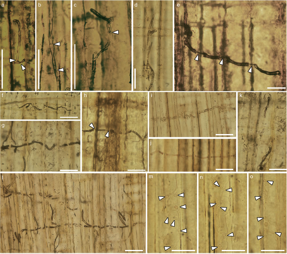
Figure 2. Blue-stain fungus in wood tissues of Xenoxylon phyllocladoides Gothan from the Jurassic of western Liaoning Province, NE China.
(a–c) Hyphae with septa (white arrow heads). (d) Hyphae growing in the cross-field zone. (e, h) Hyphae penetrating tracheid walls with appressorium-like structures and distinct hyphal pegs (white arrow heads). (f) Colonization of ray parenchyma cells by hyphae. (g, i–j, l) Hyphae horizontally penetrating the tracheid walls with appressorium-like structures. (k) Hyphae colonizing in the tracheid lumen, and passing through the bordered pits. (m–o) Slender hyphae within tracheid lumen with chlamydosporelike structures (white arrow heads).Bars: (a–d, f–n) 50 μm; (e) 25 μm.
Download: